The Computational Biology Initiative at the Center for Biomedical Informatics in collaboration with Systems Biology Department and Dana Farber Institute is developing advanced image processing and machine learning techniques to extract information about cells and cellular components from images obtained with microscope.
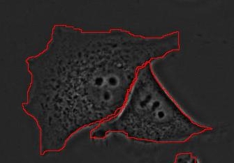
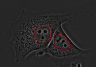
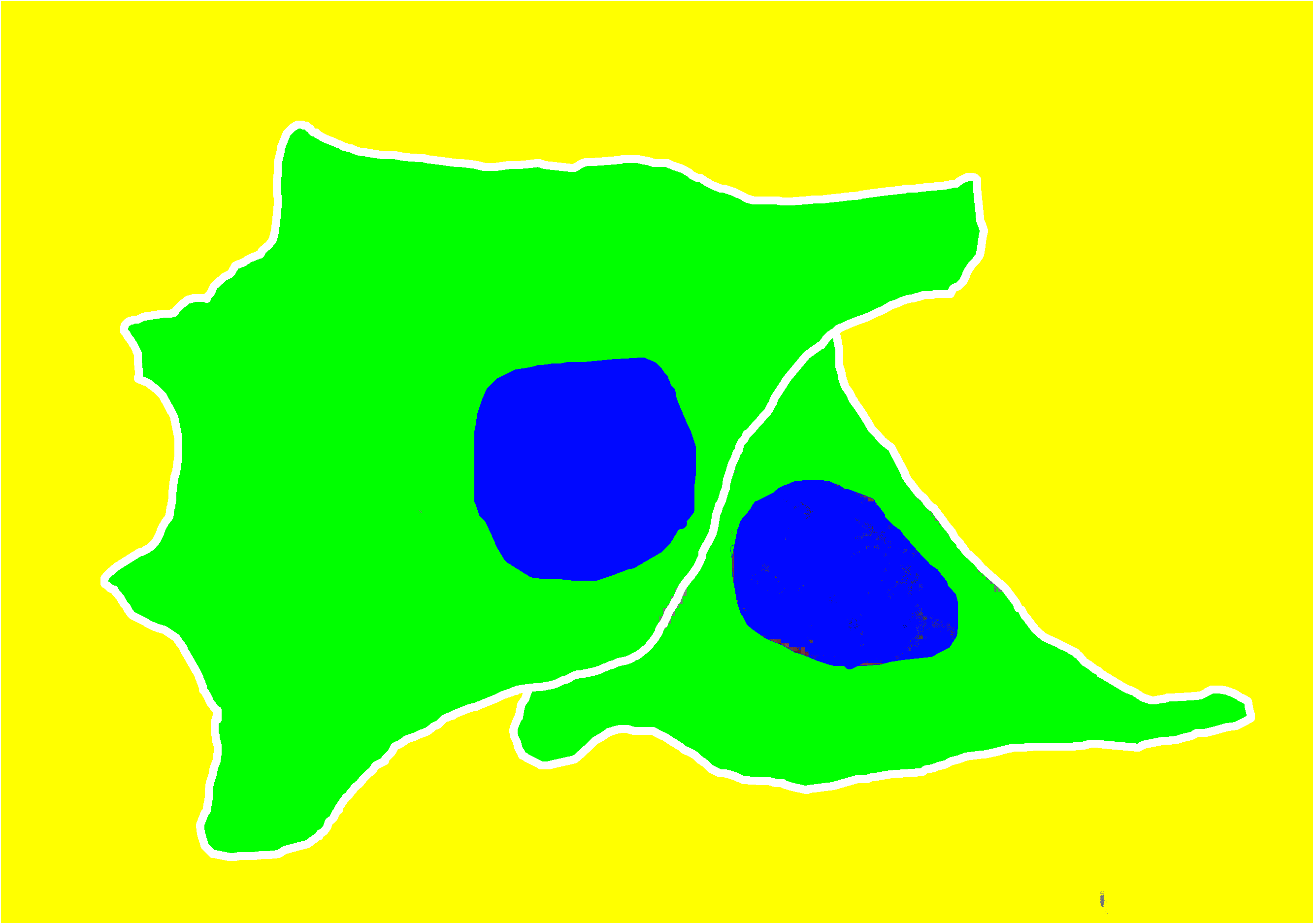
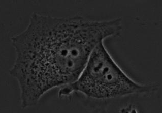 This "phase contrast" image contains two human brest cancer cells next to each other. The goal of this project is to automatically obtain segmentation so that each pixel is classified into one of three categories: part of nucleus, cytoplasm or extra-cellular space. The following images show correct outlines of the nuclear and cellular membranes as well as desired result of classification.
The project includes a wide variety of processes including: object detection, object tracking, morphological and texture feature measurement, derivative feature development and machine learning approaches for classification tasks.
Leonid Peshkin, Joaquin Goni, Alexander Loewer, Ram S. Kolluri, Galit Lahav and Dennis Wall. Morphology from Texture in Cytometry, International Journal of Tomography & Statistics 2009; 16, Issue No. S10