The Computational Biology Initiative at the Center for Biomedical Informatics (CBI at HMS) in collaboration with l Systems Biology Department and Dana Farber Institute is developing advanced image processing and machine learning techniques to extract information about cells and cellular components from images obtained via microscope.
One project is about identifying all the genes involved in cell division by quantifying the phenotype changes. This involves locating images of cells undergoing mitosis and automatically classifying these into normal and abnormal mitotic types. A picture below presents schematic representation and actual icons of (left to right) mono-polar, bi-polar and multi-polar mitosis.
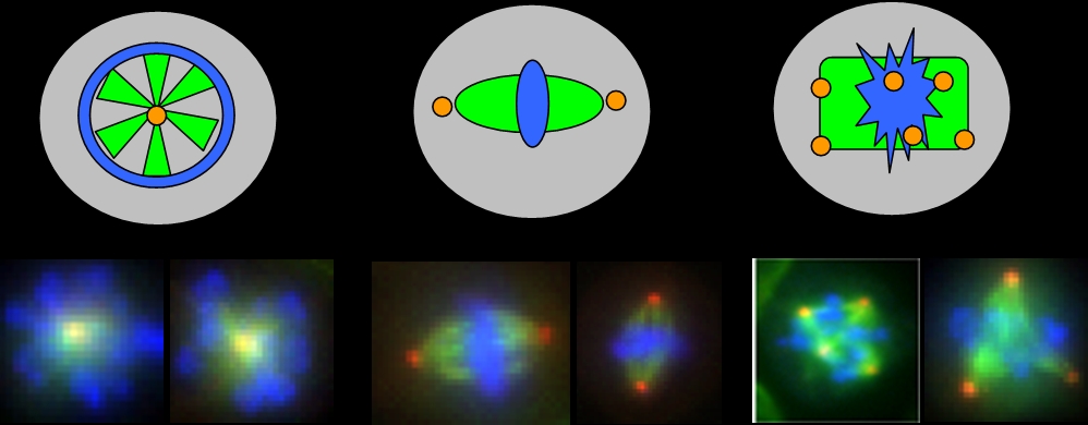 Different elements of mitotic structure are represented with blue, red and green colors respectively for chromosomes, centrosomes and spindle.
The project includes a wide variety of processes including: object detection, object tracking, morphological and texture feature measurement, derivative feature development and machine learning approaches for classification and clustering of genes affecting mitosis.
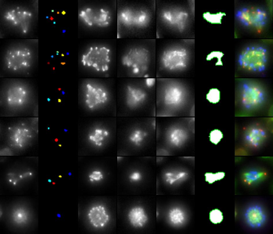
The next picture presents a gallery where each row corresponds to a single cell imaged in different fluorescent stains as well as results of centrosome and spindle detection.